The power to see inside
Our advanced scanning technology and in-depth biomarker testing to optimize your health, wellness and longevity.
Are you ready to transform your health?
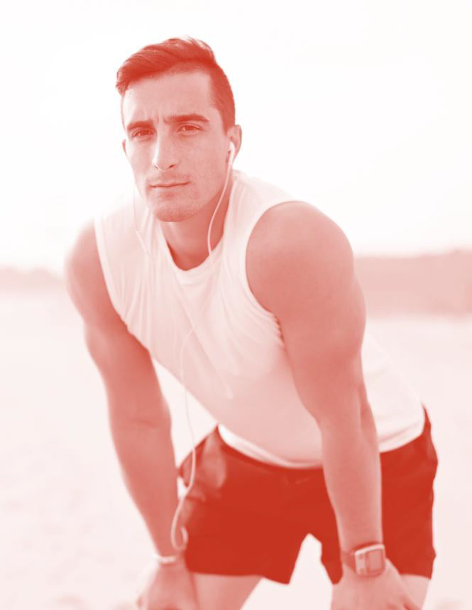
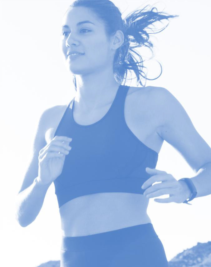
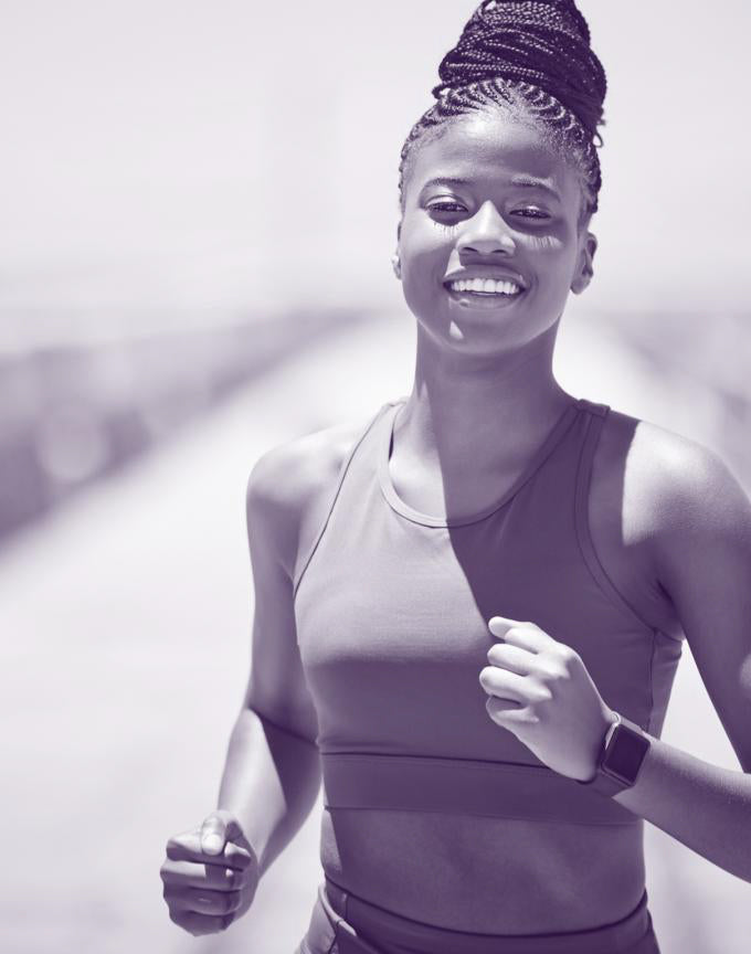
Knowing your body and seeing inside is key to transforming your health, reaching peak performance, achieving fitness goals, and living a longer, healthier life.
By embracing this journey of self-discovery, you can unlock the potential for a healthier lifestyle.

Unlock the power of your data
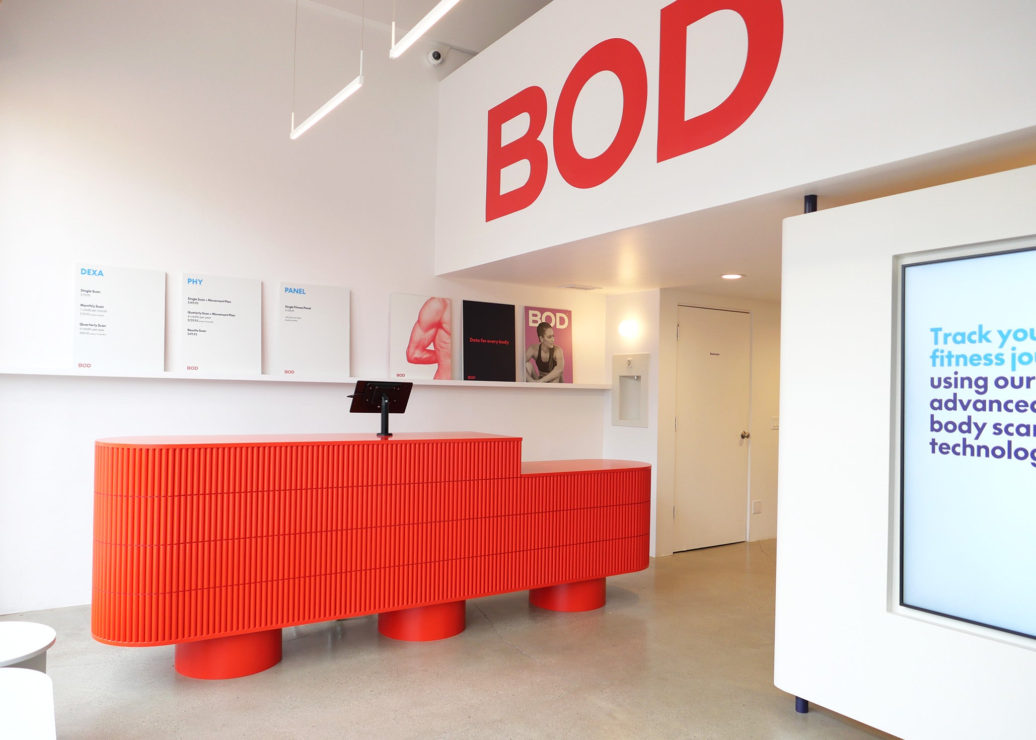
All of our four services are curated to provide a comprehensive body overview, each offering precise, insightful data and actionable recommendations.
Our advanced scanning technology and in-depth biomarker testing, provide data-driven insights to optimize your health, wellness and longevity.
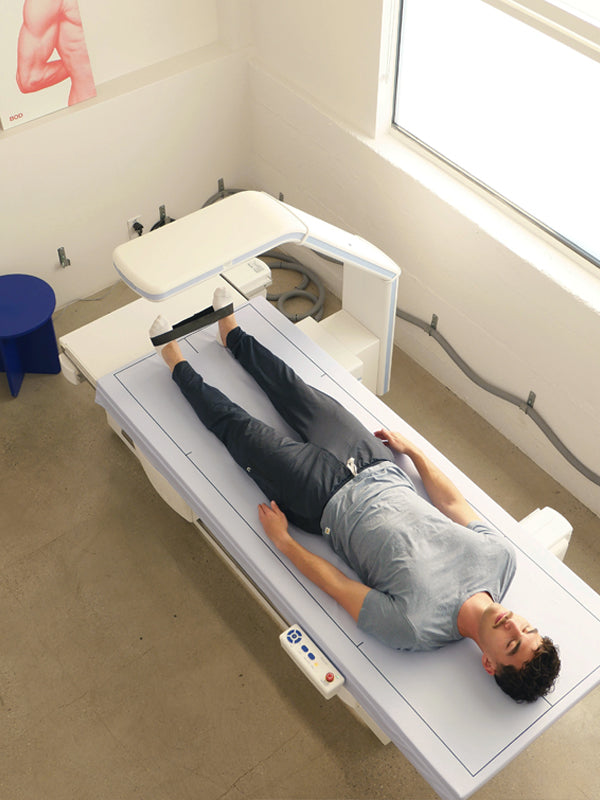
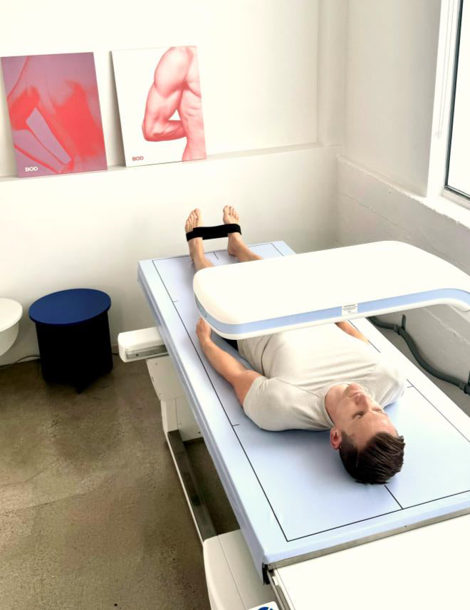
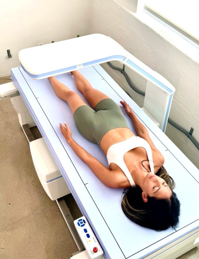
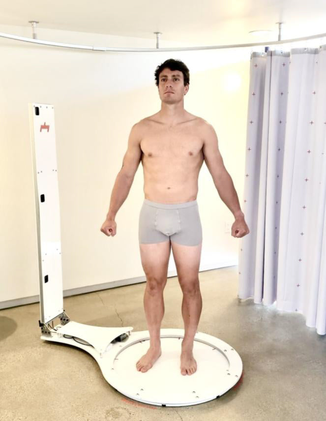
A whole-body composition DEXA scan provides a detailed analysis of your overall mass, lean mass, body fat %, visceral fat, bone density and more, which is crucial in tracking overall health and wellness.
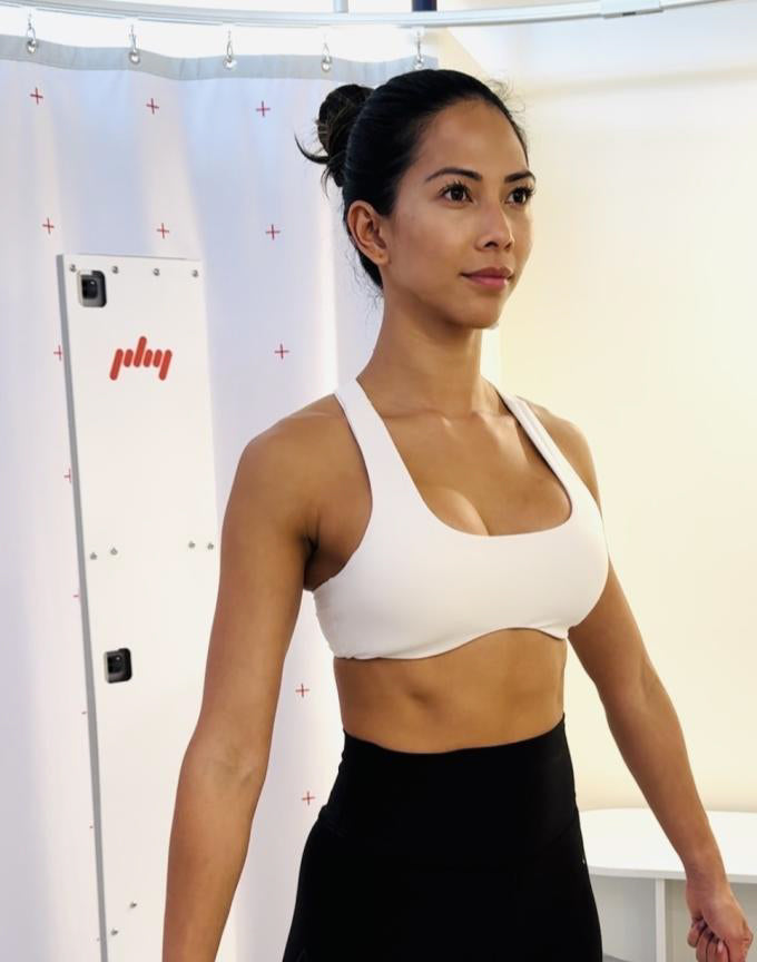
Learn moreOptimize your body alignment with a PHY scan to measure body misalignments with precision and improve mobility and flexibility. With a 6-week course of custom stretch exercises to realign the body.
Learn moreHEALTH panel
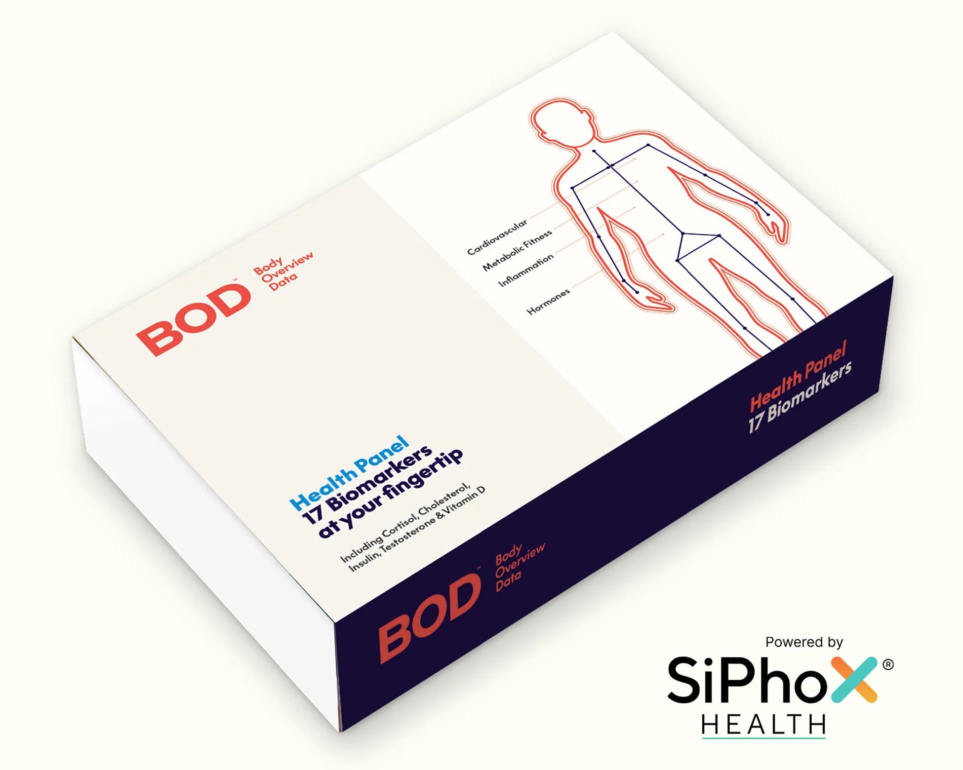
Gain key insights on 17+ health biomarkers
Get essential insights on over 17 health biomarkers with a quick finger-stick blood test.
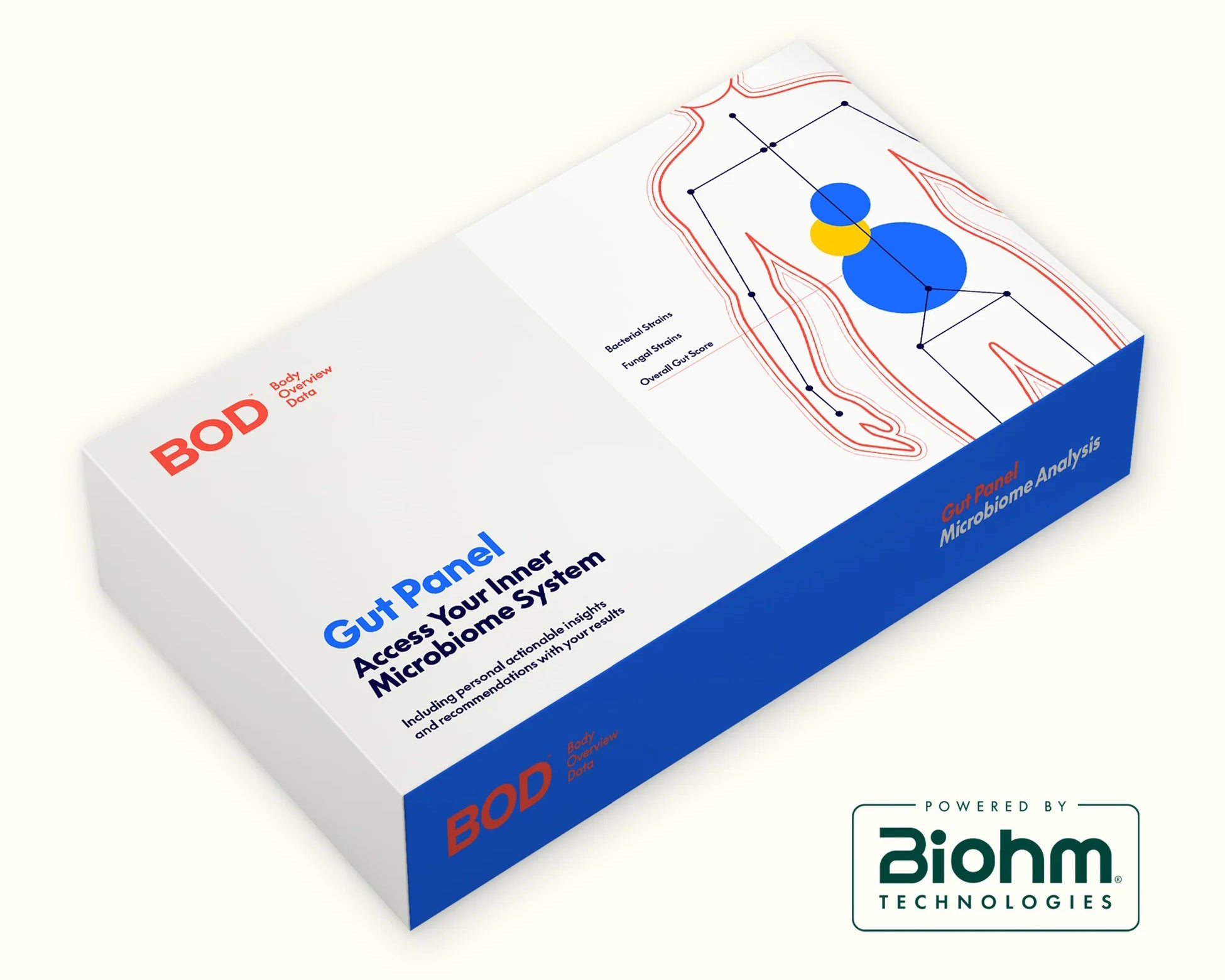
GUT panel
Access your inner microbiome system
Discover the significance of gut health for your overall wellbeing through an easy at-home sample collection.
Tracking for success
BOD isn't just a one-time health check; it's a new adoptable approach to better health.
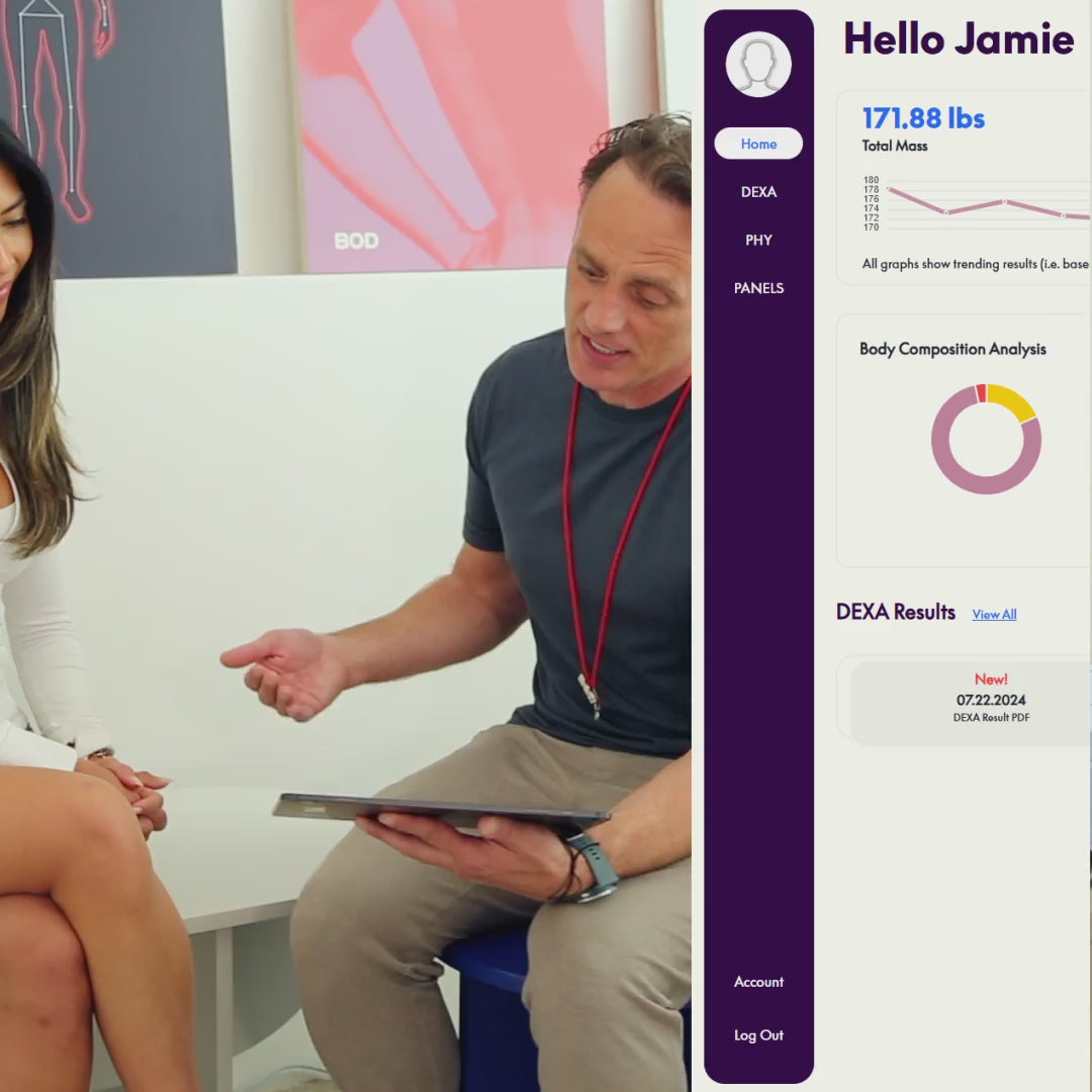
Tracking how your body changes over time, with easily accessible data in your own personal dashboard, is proven to deliver better results, secure change and drive transformation.
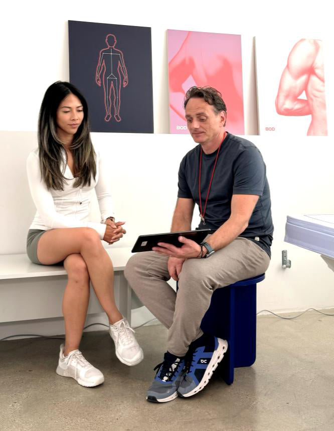
With each visit, a complimentary consultation with a BOD coach for personalized advice is included.
“Carrie and I had an incredible experience at BOD in Venice. From the moment we walked into the studio, we were impressed by the clean, modern vibe and welcoming atmosphere. The DEXA scan machine was truly first class—giving us detailed, accurate insights into our health, body composition, and fitness metrics that we had never seen before.
”

“BOD is where I go to get the critical data I need to reach my health and performance goals. I need an expert team, actionable data, and a premium environment, and that is exactly what BOD provides. A highly recommend BOD for anyone who is interested in health span, performance, and just being their best selves.
”
“I cannot recommend BOD more highly. Jeff is professional and kind, and he maintains a beautiful, clean, and tranquil space. BOD provided me with the body data and alignment assessment I was looking for, along with custom exercises and stretches to implement for improvement.
”
“I had a great experience at BOD getting both a DEXA scan and PHY scan. The customer service was outstanding—professional, friendly, and genuinely invested in making sure I got the most out of my visit. I left feeling empowered with real data to track my progress, and I’ll definitely be coming back for future scans. If you’re looking for an easy, insightful, and enjoyable experience, I highly recommend BOD.
”
“Cannot recommend BOD's location in Venice enough. Had my first DEXA scan today and left with answers to questions I didn't even know I had. Shoutout for taking the time to go over the results of my scan in detail. The experience was so informative. for those of you wishing to have a better understanding of your body composition/other biomarkers, give BOD a visit. You wont regret it.
”
“What a fantastic experience! The faculty is lovely, and the scan turned out to be very relaxing and peaceful. Most of all, the staff was amazing! Professional but also warm, they made me felt welcome and comfortable from the moment I walked in. I’ve never said this about a medical test before, but I kind of can’t wait to do it again!
”
“The DEXA Scan is so comfortable and quick. 6 minutes of meditation that I honestly wished was longer. The movement of the table is so calming. Would definitely recommend the DEXA scan. The results were extremely informative and helpful. Especially the bone density measurement for women. Katie is so kind and helpful and the facilities are beautiful.
”
“Don't wait! I only wish I did this sooner. The scan was the most relaxing 6 minutes of my day with spa-like music playing, and the environment as a whole is sparkling clean and minimalist in a way that's immediately calming. Right after the scan, they spent over 30 minutes with me, going over every detail of my report and answering all of my questions. The report itself, was incredibly comprehensive AND presented in a way that makes it perfectly clear and easy to understand.
”
“My experience at BOD was fantastic! I got a PHY scan and the process was smooth, professional, and incredibly insightful. The staff went above and beyond to make me feel comfortable, explaining everything clearly and answering all my questions
”
“I am always skeptical of
”
‘scans’ but could not have been proven more wrong by an exceptional experience at BOD. I have to shout out Katie, who saw me with great skill, humor and chemistry. She was very knowledgeable and put me at ease, guiding me through the process perfectly. The business itself is clean, sleek and aesthetic (think Apple without the queues).
“I had an amazing experience getting a DEXA scan here! The process was super thorough and comprehensive, and I left with such a clear understanding of my results. Katie was phenomenal—she took the time to explain everything in detail and provided incredibly helpful recommendations that were tailored to my needs.
”
Trusted by the worlds biggest fitness brands