May 6, 2024

The Science Behind DEXA Scans: Understanding Dual-Energy X-Ray Absorptiometry

Dual-energy X-ray absorptiometry, or DEXA, is a sophisticated medical imaging technology that measures bone mineral density and body composition. This technique uses two different X-ray beams at varying energy levels to analyze and differentiate between bone and soft tissue. This allows for highly precise measurements of bone density, as well as the fat and lean mass in different parts of the body. The DEXA scan provides comprehensive data, making it a crucial tool in medical diagnostics and health management.

body composition scanning

Fundamentals of DEXA Technology

How DEXA Utilizes Dual X-Ray Beams

DEXA technology operates on a unique principle involving two X-ray beams emitted at different energy levels. When these beams pass through the body, bone and soft tissues absorb them variably, allowing the scanner to differentiate between these components. Soft tissue absorbs the lower energy beam more, while the higher energy beam provides clearer insights into bone density. This differentiation is crucial for accurate measurements and is a key advantage of DEXA scan science over other imaging techniques.

Differentiating and Quantifying Bone, Fat, and Lean Mass

One of the standout features of advanced body fat measurement with DEXA is its ability to precisely quantify bone, fat, and lean muscle mass independently. This scan provides a detailed breakdown that includes visceral fat and total body fat percentages, which are important metabolic health indicators. For athletes or individuals in weight management programs, these metrics are invaluable for tracking progress and adjusting diet and exercise plans effectively.

Comparison with Other Body Composition Scans

While various methods are available for body composition analysis, DEXA scans are often preferred for their precision and reliability. Other commonly used techniques like bioelectrical impedance analysis (BIA) and skinfold measurements come with more limitations. BIA estimates body composition by sending a weak electric current through the body and measuring the resistance encountered, which varies according to the amount of water in the body. This method’s accuracy can be compromised by hydration levels, food intake, and other biological factors. Similarly, skinfold measurements, which involve using calipers to measure the thickness of fat at various body points, require considerable skill and experience to yield accurate results.

Accuracy and Precision of DEXA Measurements

The accuracy and precision of a DEXA scan for body composition and bone density are highly regarded. DEXA’s detailed reports help physicians diagnose and monitor conditions like osteoporosis and also allow fitness professionals to design more personalized fitness programs.

The Process of a DEXA Scan

Steps Involved

The process is quick, non-invasive, and provides critical data that can help in both diagnosis and management of various conditions. Here are the steps involved in a DEXA scan:

  1. Preparation: Before the scan begins, the patient is required to prepare adequately to ensure the accuracy of the results. They are asked to wear loose, comfortable clothing and to remove any metal objects such as belts, jewelry, or glasses, which could interfere with the X-ray images. This step is crucial as metal objects can obscure the details of the X-ray, leading to inaccurate readings or the need for repeat scans.
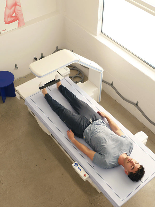
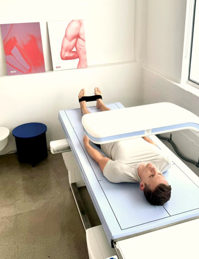
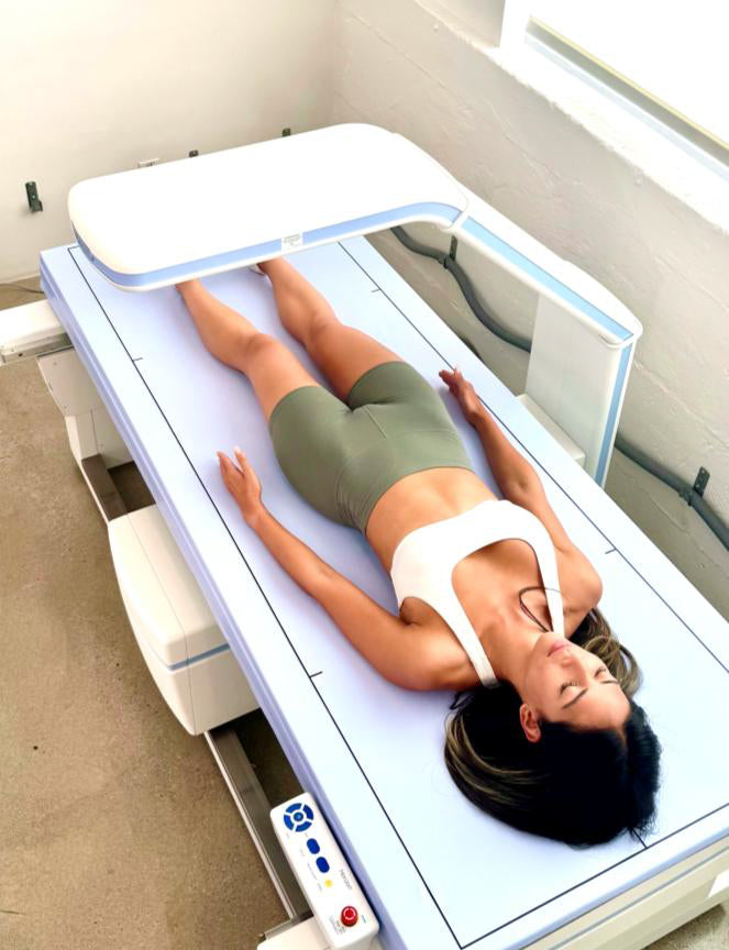
  2. Positioning: Once prepared, the patient lies flat on a padded table to ensure comfort during the procedure. Proper positioning is critical as it affects the quality of the images obtained. The scanner, which is a large, central machine, is carefully positioned over the area of interest, typically the lumbar spine and hip, which are common sites for assessing bone density. The correct positioning ensures that the scan is both effective and efficient, capturing the necessary data without the need for repositioning or additional scans.
  3. Scanning: The actual scanning process involves the machine passing over the body with low-dose X-rays at two different energy levels. This dual-energy technique allows for a more detailed analysis of the bone and soft tissues without causing any pain or discomfort to the patient. The X-rays pass through the body and are absorbed differently by bones and soft tissues, which is critical for the next steps in bone density analysis.
  4. Data Capture: As the X-rays pass through the body, they are captured by the machine’s detectors. The dual-energy beams produce detailed images that differentiate between bone and soft tissue with high precision. This data is immediately analyzed by the machine’s sophisticated software, which can distinguish subtle differences in tissue composition and bone density, crucial for accurate diagnosis.
  5. Analysis: After the scanning is complete, the machine uses the captured data to compute various metrics, such as bone mineral density and the distribution of fat and lean mass in the body. This analysis is vital for assessing the patient’s risk of osteoporosis and other conditions. The results provide a clear picture of bone health and are used to guide treatment plans and monitor disease progression or response to treatment.
  6. Report Generation: The final step in the DEXA scan process is the generation of a detailed report. This report includes both quantitative data and images obtained during the scan. It is meticulously prepared to provide a comprehensive overview that healthcare providers can use to make informed decisions about the patient’s health. The report is typically ready shortly after the scan, allowing for prompt review and discussion of the results with the patient.

The procedure is streamlined and designed for patient comfort, from preparation through to the detailed reporting of results. This allows healthcare providers to quickly and accurately assess a patient’s bone health and body composition, facilitating timely and effective medical intervention.

DEXA scan for body composition

Preparing for a DEXA Scan

To ensure the highest accuracy of DEXA scan results, patients must remain motionless during the procedure. Any movement can distort the X-ray images, leading to inaccuracies in measuring bone density and body composition. The scan typically lasts only a few minutes, during which the patient lies flat on a padded table while the scanner passes over their body. Keeping still not only enhances the precision of the results but also helps in achieving consistent data across sequential scans, which is vital for tracking changes over time. Healthcare providers often provide clear instructions beforehand to help patients understand the importance of remaining still and may offer aids like cushions or straps to minimize movement.

Patients are also advised to disclose any recent medical procedures involving barium or contrast materials to their healthcare provider before undergoing a DEXA scan. These substances, commonly used in various diagnostic tests like barium swallow studies and CT scans with contrast, can obscure the DEXA scan results. Barium and certain contrast dyes have high X-ray absorption properties, which could lead to erroneous high readings in bone density or soft tissue composition. Pregnancy is another critical factor that must be communicated to the healthcare team conducting a DEXA scan. Since the procedure involves exposure to low levels of ionizing radiation, it poses a risk to the developing fetus. Although the radiation dose from a DEXA scan is relatively low compared to other radiographic assessments, the cumulative effect of radiation exposure should be carefully managed, especially in pregnant women. Alternatives to DEXA, such as ultrasound or magnetic resonance imaging (MRI), which do not use ionizing radiation, might be recommended in these cases.

Benefits of DEXA Scans

Role in Clinical and Fitness Settings

This advanced imaging technology not only aids in disease diagnosis and treatment but also enhances personal fitness and nutritional planning. The versatility of DEXA scans in providing detailed data on bone and soft tissue composition makes it a critical tool in various settings. Here are the specific roles DEXA scans play in clinical and fitness settings:

  • Early Disease Detection: In clinical settings, DEXA scans are particularly valuable for their precision in measuring bone density. This capability allows healthcare professionals to identify early signs of bone density abnormalities, such as osteoporosis before more significant symptoms appear. By detecting these variations early, DEXA enables timely interventions that can significantly alter the course of the disease, potentially preventing severe complications and improving patient outcomes. Early detection is crucial for planning effective treatment strategies and for patient education on lifestyle adjustments and preventative measures.
  • Treatment Monitoring: These scans are essential in the ongoing monitoring of patients undergoing long-term treatments that can impact bone health, such as steroids or chemotherapy. These treatments can lead to significant changes in bone density, and regular body composition scanning provides a quantitative basis for evaluating the effectiveness of the treatment and the need for adjustments. Monitoring is essential for controlling any adverse effects, stopping the course of bone degradation, and determining the direct effect of the treatment on bone density.
  • Personalized Fitness Programs: In fitness environments, trainers rely on the comprehensive body composition analysis provided by DEXA scans to craft personalized training programs. By understanding the specific fat and muscle distribution in an individual’s body, trainers can optimize fitness regimes to focus on increasing muscle mass and reducing fat in targeted areas. This tailored approach helps in achieving more effective and sustainable fitness results, making DEXA scans a valuable tool for both trainers and clients aiming for specific health and fitness goals.
  • Nutritional Assessment: Nutritionists utilize the detailed body composition data from DEXA scans to design customized dietary plans tailored to the individual’s specific health and nutritional needs. By understanding the proportions of bone, fat, and muscle, nutritionists can recommend diets that enhance bone health, support muscle growth, and manage fat levels. Customized meal planning is essential for enhancing general health outcomes and is especially helpful for people with special dietary requirements or those going through rehabilitation.

In both medical and fitness contexts, the application of advanced DEXA scan technology helps in creating highly specific and effective strategies tailored to individual health profiles, leading to better health outcomes and optimized treatment and fitness plans.

Regional Analysis of Body Fat and Lean Mass

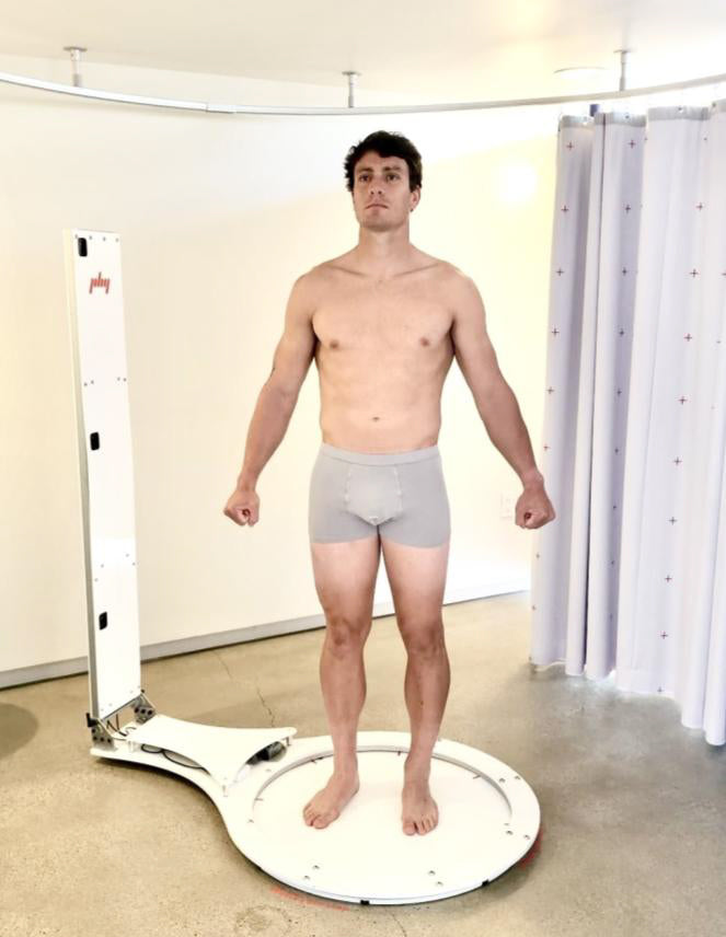
By identifying how fat and muscle are distributed across the body, specialists can better understand individual health risks associated with fat distribution patterns, such as higher risks of cardiovascular diseases linked to visceral fat. This region-specific information aids in crafting targeted strategies that can address and mitigate these risks effectively.

The Future of DEXA Scan Technology

Ongoing Advancements in DEXA Technology

As the technology continues to develop, its applications are becoming more versatile and impactful, offering greater benefits to patients and medical professionals alike. Here’s a look at the ongoing advancements:

  • Increased Resolution: Higher resolution will enable clinicians to observe finer details in bone and soft tissue structures, facilitating more precise measurements of bone density and body composition. This improvement is crucial for early detection and accurate assessment of various conditions, including subtle changes that could indicate the onset of diseases like osteoporosis. Improved resolution will also aid in more detailed monitoring of patient progress over time, particularly during treatment for bone-related ailments.
  • Faster Processing Times: Another major focus in the advancement of DEXA technology is the reduction of scan duration and data processing times. By speeding up these processes, a DEXA scan for bone density becomes more efficient, reducing the time patients spend in the machine and allowing healthcare facilities to increase the number of scans conducted daily. This enhancement not only improves the patient experience by making the procedure quicker and less cumbersome but also enhances the workflow efficiency for healthcare providers. Faster processing times mean quicker turnaround on results, enabling timely decision-making in clinical settings.
  • Enhanced Software Algorithms: Developers are working on creating more sophisticated software algorithms to analyze the data collected by DEXA scans. These advanced algorithms are designed to improve the accuracy of differentiating between various tissue types, such as distinguishing between fatty and lean tissues with greater precision. This will enhance the diagnostic capabilities of DEXA scans, allowing for more accurate assessments of body composition and bone health, which are vital for diagnosing conditions and tailoring individualized treatment plans.
  • Integration with Other Technologies: There is a significant push to integrate DEXA technology with other diagnostic tools and platforms. This integration aims to create a more holistic approach to patient health assessments, combining data from multiple sources to give a comprehensive view of a patient’s health status. By linking DEXA with other medical imaging technologies or health record systems, it can play a pivotal role in personalized medicine, helping tailor interventions and monitor outcomes in a more informed and precise manner.

These innovations not only enhance the capability of DEXA scan benefits but also position them as a cornerstone technology in the evolving landscape of medical diagnostics and personalized healthcare solutions.

bone density analysis

Future Applications and Implications

The ongoing development of DEXA technology holds significant implications for various fields, including geriatric care, sports medicine, and bariatric surgery. As the technology becomes more advanced and accessible, it could also play a key role in global health initiatives, providing critical data for large-scale health studies and public health strategies aimed at improving population-wide bone health and physical fitness.

Throughout this exploration of DEXA scan science, we’ve delved into the sophisticated nature of dual-energy X-ray absorptiometry and its profound impact on medical diagnostics and health management. The ability of DEXA scans to provide precise and detailed measurements of bone density and body composition stands out as a pillar in both clinical and fitness settings. These scans offer invaluable insights that aid in diagnosing, monitoring, and managing health conditions with a level of accuracy that few other methods can match.

Have questions?

Loading
Your message has been sent. Thank you!

To top